In this case, it shows that the "self/not self" divide is vital to our survival ...
My Thymus, Myself
[Radiolab] Today, we go to a spot that may be one of the most philosophical places in the universe: the thymus, an organ that knows what is you, and what is not you. Its mood may be existential, but its role is practical — the thymus is the biological training ground where the body learns to protect itself from outside invaders (think: bacteria, coronaviruses). But this training is not the humdrum bit of science you might expect. It’s a magical shadowland with dire consequences.
Then, we’ll leave the thymus to visit a team of doctors who are using this organ that protects you as a way to protect someone… else. Their work could change everything.
https://radiolab.org/episodes/my-thymus-myself

Also, this ...
Scientists have estimated how many ants there are on Earth. Clue: It's a lot
Ants are tiny in size but not in number. There are about 20 quadrillion ants on the Earth at any given time, a new study has estimated. That's 20,000 trillion individuals.
The estimate is two to 20 times higher than previous ones, according to the study, published in the journal Proceedings of the National Academy of Sciences on Monday.
https://edition.cnn.com/2022/09/20/w...scn/index.html
... as much as, inwardly, every t-cell and the thymus of your body is you ... and you are the thymus youing ...
... and you are dinosaur fossils too dinosaur fossiling ...

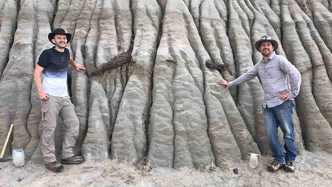
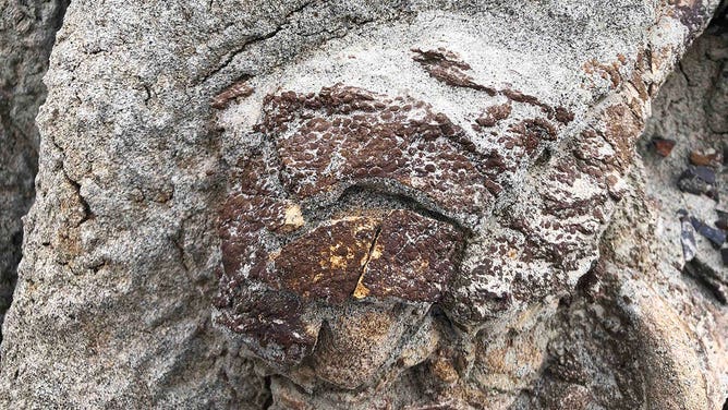
Scientists think they’ve found a rare ‘dinosaur mummy’ in Canada
... A so-called "dinosaur mummy" may be contained within a rocky hill in Canada, according to scientists.
In a statement issued earlier this month by the University of Reading, this rare type of fossil refers to finds where the entire skeleton of the dinosaur may be preserved.A so-called "dinosaur mummy" may be contained within a rocky hill in Canada, according to scientists. ... According to scientists, the exposed parts of the fossil, which include the animal’s tail and right hind foot, indicate it was likely a hadrosaur – a large, duck-billed, herbivorous dinosaur. ... What makes this find even more unique is that exposed parts of the fossil appear to be covered in fossilized skin, according to Caleb Brown, a Ph.D. at the Royal Tyrrell Museum, which will take possession of the find once it is excavated.
"This suggests that there may be even more preserved skin within the rock, which can give us further insight into what the hadrosaur looked like," Brown said.
https://news.yahoo.com/scientists-th...204959750.html
Gassho, J
stlah


")


Leave a comment: